- Share
- Share on Facebook
- Share on LinkedIn
Nanochemical Imaging and Metallobiology of Biologic Systems
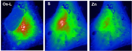
The synchrotron micro- and nano-probe spectroscopic techniques in continuous development and progress allow a unique mesoscale capability to explore and elucidate the distribution, concentration and chemical state of metals inside tissues and cells down to the organelle level. This contribution is not only highly challenging but represents important objectives in modern analytical chemistry and an essential step towards the precise understanding of some cellular physiopathological or toxicological processes.
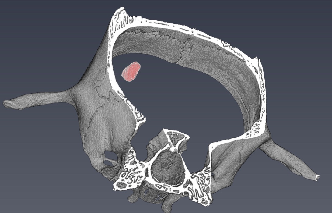
OsteoArticular Diseases
Osteoarticular diseases are the most prevalent chronic pains and long-term disabilities with hundreds of millions of people affected worldwide. Until now there is a lack of imaging modalities for performing long-term follow-ups of osteoarticular diseases in order to understand their mechanisms or to prove the efficiency of therapy on small animals. In this axis, we propose to develop X-ray Phase Contrast Imaging that presents the great advantage of being sensitive to the different tissues constituting a joint.
Imaging Lung Structure and Function
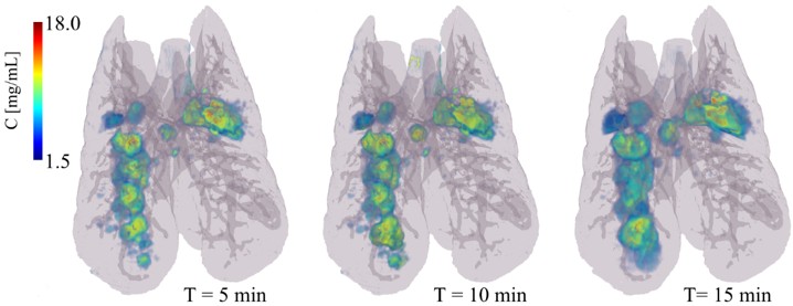
The uneven distribution of ventilation has major fundamental and clinical implications. Ventilation heterogeneity affects the matching of regional ventilation and perfusion leading to less efficient gas exchange, and can significantly affect the apparent degree of mechanical obstruction. Synchrotron imaging of regional lung structure and function takes full advantage of the high flux and energy-tunability of synchrotron radiation and is applied to the pathophysiological investigation of lung diseases such as asthma and ventilator-induced lung injury
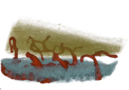
Regenerative Medecine
Recent progress in the field of regenerative medicine has raised hopes for a treatment breakthrough in a wide range of debilitating conditions. Stem cell therapies are currently evaluated in many clinical trials. The core objective of this project is to develop the imaging methods to monitor during several weeks the fate of stem cells on one hand and of cell-embedding hydrogels on the other hand, after in-situ transplantation in rodent models of ischemic stroke. We combine both spectral approaches and phase contrast imaging at the synchrotron and we compare the obtained results on the Spectral Photon CT scan available in Lyon.
- Share
- Share on Facebook
- Share on LinkedIn