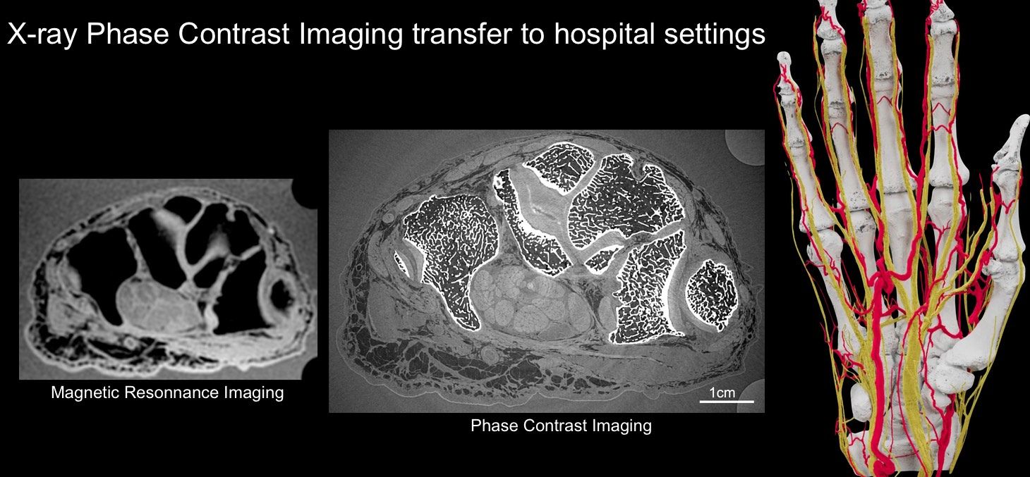
X-ray Phase Contrast Imaging
X-ray Phase Contrast Imaging is sensitive to the different tissues constituting a joint. Find out more on how we propose to transfer this technology to hospital settings.
Director's words
"We develop new technologies and methods applying radiation from high-energy synchrotron sources to the biomedical sciences. We offer a platform of diverse synchrotron X-ray technologies ranging from multimodal quantitative microscopy to macroscopic in vivo and functional imaging, as well as radiation therapy and radiosurgical approaches, both in preclinical models and clinical trials. The lab is currently located at the European Synchrotron Radiation Facility and will be moving to the Université Grenoble Alpes Campus in 2021".
Partnership
STROBE is a member of Labex PRIMES, France Life Imaging, and has many national and international partners.
![]()
Recent Publication
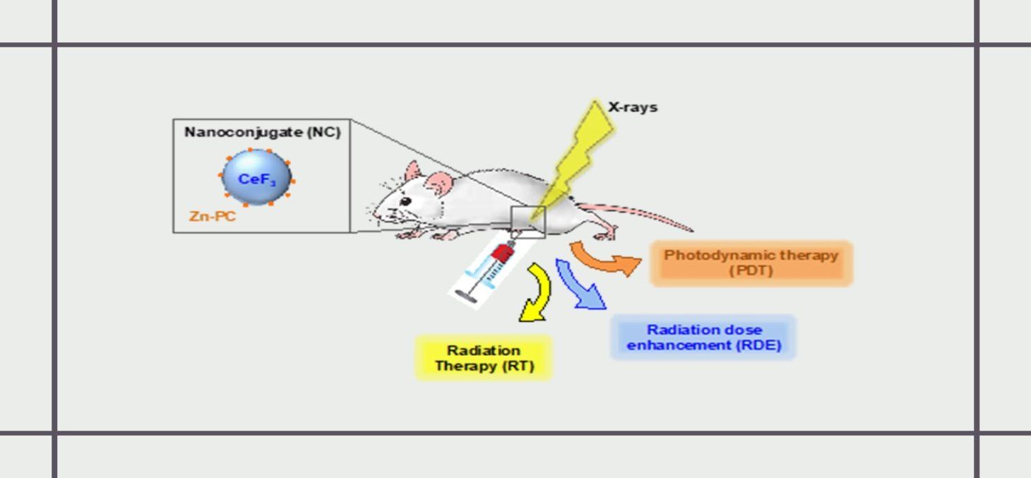
Radiation Dose‐Enhancement Is a Potent Radiotherapeutic Effect of Rare‐Earth Composite Nanoscintillators in Preclinical Models of Glioblastoma.
The photodynamic therapy (PDT) remains limited by the low penetration of light in tissues. In order to overcome this restriction, it has been proposed to conjugate the photosensitizers on to nanoscintillators. Those down-converting nanoparticles absorb X-ray and convert those penetrating radiations into visible light that can subsequently excite the photosensitizer and induce PDT independently of the tumour location. This concept of radiation therapy/deep tissue PDT upon synchrotron radiation applied to an aggressive syngeneic model of orthotopic glioblastoma has been published recently in Advanced Science.
FRM speak about us
Cancers : vers une radiothérapie innovante contre les glioblastomes.
BIRS Bioengineering School 2026

April 13-17, 2026 Villa del Grumello, Como – Italy