- Share
- Share on Facebook
- Share on LinkedIn
Current Status and limitations
Current clinical and preclinical imaging modalities have limitations in the detection of early cartilage and bony changes despite their crucial roles toward the development of therapeutic options of cartilage diseases. Indeed, conventional X-ray absorption-based Computed Tomography (CT) allows a clear 3D visualization of bone tissues but provides reduced sensitivity to the soft tissues. Changes in the composition of joint cartilage or soft tissues are usually rather evaluated using Magnetic Resonance Imaging (MRI). Yet, the images acquired by MRI struggle to render properly the bony changes and the micro-calcifications.
Phase contrast imaging: a new modality for osteoarticular diseases
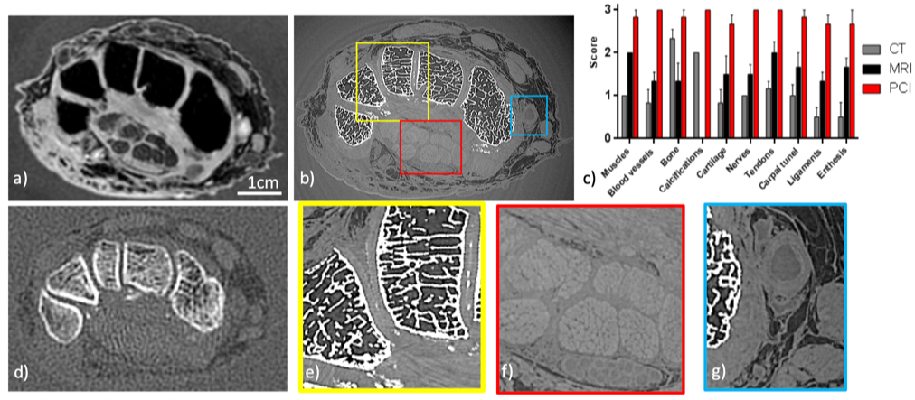
Since the seminal work of Roentgen, X-ray imaging is based on the same physical phenomenon: the absorption of light by the tissues. But refraction, the fact that light is deviated when passing through matter, is more preponderant than absorption. Indeed, the refraction index of materials can be a thousand times greater than its counterpart absorption factor for light elements. This translates into a much greater contrast for soft tissues with X-ray imaging methods based on the sensing of the phase (so-called Phase Contrast Imaging (PCI)) in comparison to the conventional method based on absorption. The figure below is extracted from a recent study in which we evaluate the diagnosis potential of PCI-CT for joint characterization. In this study, X-ray PCI was performed on three cadaveric human hands and wrists using a synchrotron monochromatic beam. Conventional CT, MRI and US exams were also performed on these three samples using routine procedures as well as research protocols. Six radiologists and rheumatologists from Grenoble University Hospital independently evaluated qualitatively and semi-quantitatively the images’ quality.
Noise Based Imaging: a simple set-up for PCI and X-ray functional imaging
Noise in images is generally discarded because in general it is thought that it obscures the signal, degrades the signal and does not add any information. Indeed, since the early days of imaging there is a continuous effort to reduce the level of noise as low as possible. However, in wave physics and especially, seismology, scientists developed some tools known as “noise correlation” to extract useful information. In this project, we propose to apply such a methodology in X-ray imaging. By adding a controlled generator of noise during the acquisition, Noise Based Imaging (NBI) is an imaging methodology to acquire new types of information in X-ray images. Thanks to image processing methods it is indeed possible to track down sample properties such as its refraction, its absorption, its scattering or its mechanical properties.
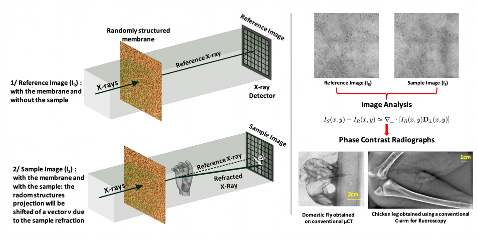
The experimental set-up involves only the sample, an X-ray imaging detector and a randomly structured membrane containing random high spatial frequency structures of a few pixels size that will create a static noise or in other words a random modulation. This set-up besides its simplicity of implementation has the main advantages of having no field of view limitation other than the detector (the diffusor is easy to manufacture), no resolution limitation other than the optical system (noise of a few pixels can be generated down to tens of nm) and finally the requirements on the beam coherence are low. NBI is radiation dose efficient for two reasons: i) no absorbing element is used between the sample and the detector meaning that all photons passing through the sample are eventually contributing to the image formation; ii) only a single couple of images (with and without the sample) has to be acquired unlike most of the GBI or EI methods.
Noise Based Imaging
Responsible: Emmanuel Brun
People involved: Ludovic Broche, Laurene Quenot
Collaborators: Dr A. Mirone, Prof David M. Paganin, Prof K. Pavlov,
Alumni: Helene Rougé-Labriet
- Share
- Share on Facebook
- Share on LinkedIn