- Share
- Share on Facebook
- Share on Twitter
- Share on LinkedIn
- Share url
It is thus essential to develop imaging modalities that allow the monitoring of the administered cells in order to follow their fate in a non-invasive manner. Labeling the injected cells using gold nanoparticles (AuNPs) allows monitoring the cells with CT-based imaging techniques.
Novel experimental CT techniques, namely Phase Contrast [1] and Spectral Imaging [2], that have been introduced in synchrotrons in the past years and recently translated to the clinics[3], [4], may help to overcome those limitations. While they were developed in parallel, the aim of this project is to combine the two approaches for improved segmentation of labeled-cells with a focus on small animal follow-up with a model of focal cerebral injury mimicking stroke infarct and an osteoarthritis model.
This project, named BREAKTHRU, is funded by the French Research National Agency (ANR-18-CE19-003-01).
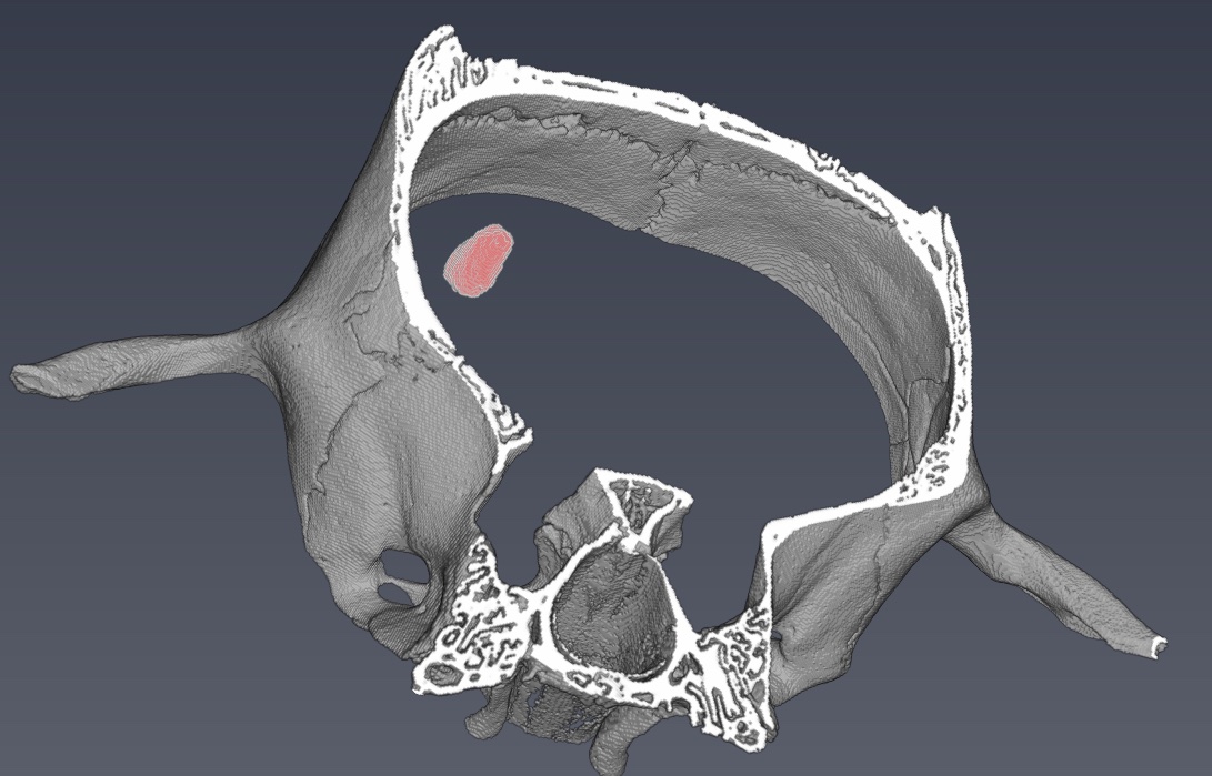
The figure below shows preliminary results
References
- R. Ashton, J. L. West, and C. T. Badea, “In vivo small animal micro-CT using nanoparticle contrast agents,” Front. Pharmacol., vol. 6, p. 256, Nov. 2015, doi: 10.3389/fphar.2015.00256.
- A. Bravin, P. Coan, and P. Suortti, “X-ray phase-contrast imaging: from pre-clinical applications towards clinics.,” Phys. Med. Biol., vol. 58, no. 1, pp. R1–R35, Jan. 2013, doi: 10.1088/0031-9155/58/1/R1.
- W. Thomlinson, H. Elleaume, L. Porra, and P. Suortti, “K-edge subtraction synchrotron X-ray imaging in bio-medical research,” Phys. Medica, vol. 49, pp. 58–76, May 2018, doi: 10.1016/j.ejmp.2018.04.389.
- K. Willer et al., “X-ray dark-field imaging of the human lung—A feasibility study on a deceased body,” PLoS One, vol. 13, no. 9, p. e0204565, Sep. 2018, doi: 10.1371/journal.pone.0204565.
BREAKTHRU project
People Involved:
Helene Elleaume
Marlene Wiart (Carmen)
Emmanuel Brun
Clément Tavakoli
Partners:
Carmen
Institute for Regenerative Medicine & Biotherapy
Grenoble Institute of Neurosciences
CERMAV
UPenn
- Share
- Share on Facebook
- Share on Twitter
- Share on LinkedIn
- Share url